Blog
Why Choose Digital X-ray Equipment for Dental Safety?

In an era where medical radiation exposure is under intense scrutiny, dental practices face a critical challenge: nearly 15% of patients express concerns about X-ray radiation during routine visits. Traditional dental radiography, while essential for diagnosis, has long been associated with radiation exposure risks that worry both patients and healthcare providers. lemaclinic.com Digital X-ray equipment emerges as a revolutionary solution, fundamentally transforming how dental practices balance diagnostic necessity with patient safety. This advanced technology not only reduces radiation exposure by up to 90% but also delivers superior image quality for more accurate diagnoses. As dental practices navigate the evolving landscape of patient care and safety regulations, understanding the implementation and benefits of digital X-ray systems becomes paramount. This article explores how digital radiography is reshaping dental safety protocols while enhancing diagnostic capabilities, providing practical insights for dental professionals considering this crucial technological upgrade.
The Critical Shift from Traditional to Digital Dental X-ray Machines
Traditional film-based dental X-ray systems have long been the backbone of dental diagnostics, but their limitations have become increasingly apparent in modern practice. These systems require chemical processing that generates hazardous waste, with each film development releasing silver-containing solutions that harm the environment. The workflow inherently demands multiple steps – from precise exposure timing to manual development – creating opportunities for errors and retakes. Traditional systems also lack the ability to adjust image quality post-capture, often resulting in repeated exposures that increase patient radiation dose.
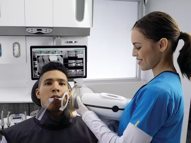
Digital radiography marks a revolutionary advancement, delivering up to 90% reduction in radiation exposure compared to conventional film. This transformation began with the introduction of digital sensors in the 1990s and has evolved through continuous technological improvements in sensor sensitivity and processing capabilities. Modern dental X-ray machine eliminate chemical processing entirely, while enabling instant image acquisition and real-time quality assessment. The transition represents not just an upgrade in technology, but a fundamental shift in operational efficiency, environmental responsibility, and patient care standards. With digital systems now capable of producing diagnostic-quality images at a fraction of the radiation dose, the move away from traditional film-based systems has become not just advantageous but essential for forward-thinking dental practices.
How Digital X-ray Equipment Enhances Diagnostic Accuracy
Superior Imaging Capabilities of Advanced Dental Imaging
Digital X-ray systems revolutionize dental diagnostics through unprecedented image clarity and manipulation capabilities. Modern sensors capture images with up to 65,536 grayscale levels compared to film’s 25 shades, enabling dentists to detect subtle variations in tooth structure and bone density. Advanced image enhancement tools allow real-time adjustments of contrast and brightness, revealing hidden details that might otherwise go unnoticed. The ability to magnify images up to 300% without quality loss proves invaluable for detecting microfractures and early-stage cavities, while split-screen comparison features enable precise monitoring of dental conditions over time.
AI Integration and Diagnostic Consistency
Artificial intelligence algorithms now augment dental diagnostics by automatically flagging potential abnormalities, reducing the risk of missed diagnoses. These systems analyze thousands of reference images to identify patterns indicating cavities, periodontal disease, and potential malignancies with remarkable accuracy. The standardization provided by AI-assisted diagnosis ensures consistent evaluation across different operators and appointments, minimizing subjective interpretation variations. Advanced comparison tools track subtle changes in dental structures across multiple visits, creating a comprehensive timeline of oral health that aids in early intervention and treatment planning. This integration of AI technology with digital imaging represents a significant leap forward in diagnostic precision and patient care quality.
Radiation Reduction: Digital X-ray’s Impact on Patient Safety in Dentistry
Scientific evidence consistently demonstrates that digital X-ray systems reduce radiation exposure by 70-90% compared to traditional film-based methods. This dramatic reduction stems from advanced sensor technology that requires significantly less radiation to produce diagnostic-quality images. Digital sensors utilize highly sensitive CMOS or CCD technology that can detect and process X-ray photons more efficiently than conventional film, enabling shorter exposure times while maintaining excellent image quality. For pediatric patients, this reduction is particularly crucial, as developing tissues are more susceptible to radiation effects. Digital systems allow for child-specific protocols that further minimize exposure while maintaining diagnostic accuracy.
Pregnancy safety protocols benefit substantially from digital radiography’s reduced radiation requirements. When X-rays are necessary during pregnancy, digital systems enable ultra-low dose protocols that maintain diagnostic value while minimizing fetal exposure. For patients requiring frequent dental visits, such as those undergoing extensive restoration work or orthodontic treatment, the cumulative benefit of reduced radiation exposure becomes increasingly significant. Modern digital systems strictly adhere to the ALARA (As Low As Reasonably Achievable) principle through built-in exposure optimization features that automatically adjust settings based on the specific diagnostic requirements, patient characteristics, and anatomical region being examined. This intelligent dose management ensures that radiation exposure is minimized without compromising the diagnostic quality necessary for accurate treatment planning.
Selecting Reliable Digital X-ray Equipment and Dentist Equipment Suppliers
Technical Evaluation Criteria
When evaluating digital X-ray systems, sensor technology selection proves crucial for long-term performance. Leading manufacturers like fsroson offer CCD sensors with superior image quality and durability but come at a higher initial cost, while CMOS sensors provide excellent value with comparable image quality and lower power consumption. Integration capabilities with existing practice management software determine workflow efficiency, making it essential to verify compatibility with current systems and data transfer protocols. Future-proofing considerations should include upgrade paths for sensor technology, software updates, and expanding storage capacity to accommodate growing image databases. Look for systems offering DICOM compliance and open-architecture solutions that allow seamless integration with emerging technologies.
Vetting Dentist Equipment Suppliers
Supplier evaluation begins with verification of FDA clearance and CE marking for all equipment components. Examine service level agreements for response time guarantees, preventive maintenance schedules, and emergency support availability. Training programs should include comprehensive initial staff certification, ongoing education modules, and access to online resources. Compare warranty terms beyond standard coverage periods, focusing on sensor replacement policies, software updates, and hardware maintenance. Request references from existing clients, particularly practices of similar size and scope, to assess supplier reliability and post-installation support quality. Evaluate financial stability and market presence to ensure long-term viability of support and upgrades.
Implementation Roadmap: Transitioning to Digital X-ray Systems
Step 1: Clinic Readiness Assessment
Successful digital X-ray implementation begins with a comprehensive clinic evaluation. Dedicated space requirements include minimum clearances of 4×6 feet for sensor storage, workstation setup, and equipment positioning. Electrical infrastructure must support consistent power delivery through dedicated circuits and surge protection. Network capabilities require minimum 1 Gbps ethernet connectivity or enterprise-grade Wi-Fi for image transfer. Staff competency mapping should identify existing digital technology proficiency levels and establish baseline training requirements for each team member.
Step 2: Phased Installation Protocol
The integration process follows a staged approach to minimize disruption. Initial sensor deployment focuses on high-volume operatories while maintaining backup film capability. Software installation encompasses practice management integration, image storage configuration, and automated backup systems. Critical checkpoints include network security protocols, data encryption setup, and disaster recovery planning with cloud-based redundancy.
Step 3: Staff Training and Compliance
Personnel development encompasses three key areas: technical proficiency, safety protocols, and patient communication. Radiation safety certification updates include digital-specific exposure guidelines and positioning techniques. Workflow integration workshops provide hands-on experience with new imaging software and quality control procedures. Patient education materials should be developed to explain reduced radiation benefits and enhanced diagnostic capabilities, ensuring clear communication about the improved safety standards digital systems provide.
The Future of Safe and Efficient Dental Diagnostics
The transition to digital X-ray equipment represents a defining moment in modern dentistry, where enhanced diagnostic capabilities align perfectly with elevated safety standards. By reducing radiation exposure by up to 90% while delivering superior image quality, digital systems effectively address the longstanding concern of radiation safety in dental diagnostics. The integration of AI-driven analysis tools, combined with advanced imaging capabilities, establishes a new benchmark for diagnostic accuracy and patient care. For dental practices considering this transition, the initial investment is justified by long-term benefits: improved workflow efficiency, reduced environmental impact, and enhanced patient confidence. As dental imaging technology continues to evolve, early adopters of digital X-ray systems position themselves at the forefront of safe, efficient, and patient-centric care. The future of dental diagnostics lies in these intelligent, safety-conscious systems that protect both patients and practitioners while delivering unprecedented diagnostic precision. Making the switch to digital X-ray equipment isn’t just an upgrade in technology—it’s an investment in the future of dental safety and care quality.
